
Some of you will feel like you’ve been poked, prodded and scanned within an inch of your life between from finding your initial symptom, throughout diagnosis and treatment and beyond. Others will just have a few initial scans and that might be it. It depends on your pathology, how straightforward it is, and on your medical team.
I’ve just briefly described a few of the most common tests, scans and x-rays used for a cancer diagnosis below. All include links to more in depth descriptions if needed.

Blood Tests. These will be the most regular tests you will have if you are having chemotherapy. I had to get over my fear of needles and blood pretty quickly. (Actually, not sure I am over it – still makes me go weak at the knees and feel sick!). You will have your blood tested a couple of days before every infusion to ensure that your blood count and immune system are strong enough to cope with the next chemo. Some of you may also have regular blood tests following diagnosis, throughout treatment and once treatment has finished to monitor your tumour markers. Certain chemicals are released into the bloodstream when you have cancer and these can be measured to diagnose a cancer and then monitored to see how you respond to treatment and whether there is evidence of it having returned. However, most hospitals in the UK don’t seem to monitor tumour markers as it seems they are not 100% reliable and can lead to false positives.

Mammogram. This machine squishes your poor boobies inbetween plates in order to take an x-ray of them. It can be slightly uncomfortable – particularly if you are young and have firm breasts. This form of scanning does include radiation.
 Ultrasound. Easy peasy. The radiographer will squeeze some gel onto your skin and slide the ultrasound transducer around to get a full image of your breast and/or lymph nodes. This can be to simply see what’s going on inside or as a biopsy guide. No radiation is involved – so this is perfectly safe.
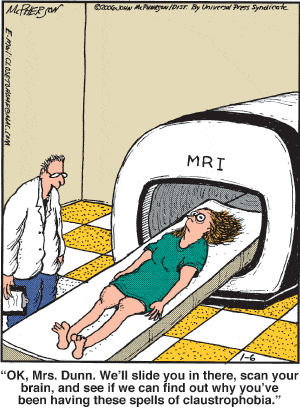
MRI. (Magnetic Resonance Imaging). This is the claustrophobic one that people get worried about – but it’s not that bad, so please don’t worry. I had an MRI after my diagnosis to get a better idea of the size and extent of the tumour and lymph node involvement as it didn’t show up on an ultrasound or mammogram. I also had one following chemo to see if the tumour had shrunk, and another to check my spine when I was suffering from severe back pain.
You will be given instructions beforehand regarding eating and drinking. You will probably be injected with a contrast dye to show up the tissue better. You will need to remove all jewellery and anything metal (due to it working with magnets) and will need to sign paperwork to say that you have no metalwork or metal fragments in your body. For breast cancer diagnostics, you are then positioned on the bed – face-down with your boobs in a couple of holes for the first scans and then again on your back – and slowly enter the tube. They might offer earphones with a selection of music and you will be given an alarm to hold in one of your hands to press in emergencies. There is also an intercom and the radiographer should check in with you at regular intervals during the scan. You have to lie VERY STILL (the images are blurred by movement) for reasonably long periods of time (up to about 40 minutes). It is painless and harmless (no radiation is involved) but it does make a bloody loud noise! Lots of whirring and clinking and clanking and banging. I just lie very still with my eyes closed, listen to the music and try and imagine myself somewhere else. It’s the noise and the enclosed space that you have to get your head around…but it’s really not that bad. If you do suffer from claustrophobia tell your consultant or the radiographer in advance. They can give you a sedative if needed or just keep a close eye on you and talk to you more regularly during the scan. If you were to panic they would get you out in a matter of seconds.

CT scan. (or CAT scan – Computerised (Axial) Tomography). These scans are very quick and painless and give a detailed picture of your insides. I had a CT scan after my diagnosis to check for spread and then another for my radiotherapy planning (see Z is for… Zapping those Cancer Cells with Radiotherapy for my experience with radiotherapy planning). Depending on what part they’re scanning, you might be asked not to eat that day, drink lots of water, and/or you may be injected with a contrast dye. You will be sent the instructions in advance. I had to strip to my knickers and don a hospital gown and lie on the bed which glides in and out of a small doughnut shaped hole. You are not enclosed, so there are no claustrophobic feelings as with the MRI scan – and it is really quick and painless. Radiation is involved.

X-ray. Another very quick, easy and painless way of checking what’s going on inside you. Radiation is involved, but your consultants will be bearing this in mind when weighing up the benefit versus the risk. The radiographer will put you in the correct position and then stand behind a screen while they take the pictures. All you need to do is stay still for a few seconds.

PET scan. (Positron Emission Tomography). These scans are to see if the cancer has spread to other parts of your body. You are asked not to eat anything on the day of the scan. When you arrive you are injected with a radioactive drug after which you have to lie down for an hour while you wait for the radioactive juice to spread around your body. You are then put through the scanner which can also take up to an hour. This is more like the CT machine than an MRI – so again, you don’t have the claustrophobic feeling, but radiation is involved and it is quite a lengthy process – but completely painless.
 ECG. (Electrocardiogram). You will probably be given one of these before you start chemo to check that your heart will cope with the toxic drugs. Again, very quick and easy with no radiation involved. You will have some sticky patches called electrodes stuck to various parts of your chest, arms and legs. You feel nothing and it is harmless. It then picks up the electric signals of your heartbeat which is used to check that your ticker is tickety-boo.

ECHO. (Echocardiogram). If you are having Herceptin you will also have your heart regularly monitored as it can seriously affect the heart (as it did in my case – I had to stop Herceptin 5 months after having started. It isn’t common, but it happens, so it is important to monitor you closely). It’s a painless and harmless procedure with no radiation involved- rather like the ultrasound. You have a blob of gel on your chest and the consultant will slide the transducer around, taking various measurements and occasionally listening to the whoosh of your heart.

MUGA Scan. (Multiple-Gated Acquisition Scan). This is another heart scan, used for drugs like Herceptin. You might be asked to fast for a period beforehand. Electrodes will be stuck to your chest and you will be injected with a radioactive tracer. You then lie still on a table while a special camera uses gamma rays to track the tracer and see how well blood is pumping through your body and how strong your heart is. This is a lengthier process than an ECG or ECHO and can take up to a couple of hours, does include radiation, but is completely painless (apart from the quick injection at the start if you’re not keen on needles!).


Leave a comment